


-
-
Overview
-
BI is a DFHBI (3, 5-difluoro-4-hydroxybenzylidene imidazolinone) derivative that binds Broccoli™ with higher affinity and deliver improved cellular fluorescence. In cells, BI has been found to stabilize the Broccoli™ structure and promote its folding. Additionally, Broccoli™/BI complex is significantly more photostable due to impaired light-induced photoisomerization, and rapid unbinding of photoisomerized cis-BI. BI is cell-permeable with negligible toxicity in living cells and can be used to label any genetically encoded Broccoli™ RNA tag without disrupting biological functions. Importantly, the optimized fluorescence properties of BI enable live single-molecule imaging of Broccoli™-tagged mRNA transcripts in mammalian cells. Compared to MS2-tagged mRNA, Broccoli™-tagged mRNA puncta exhibit similar size and puncta diameters fall within a single Gaussian curve.
Data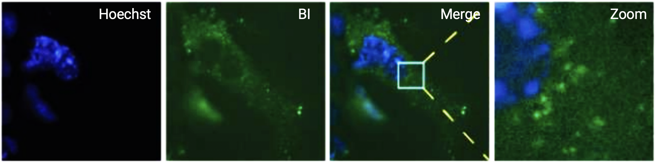
Figure 1. Live-cell imaging of COS7 cells expressing β-actin mRNA transcripts tagged with 24 copies of Broccoli™ in the 3’UTR. Cells were incubated with 0.5 µg/ml Hoechst 33342 stain (blue) and 10 µM BI (green) and images were acquired using a 500 msec exposure with a GFP filter set. Mobile fluorescence puncta were observed only when Broccoli-expressing cells were treated with BI.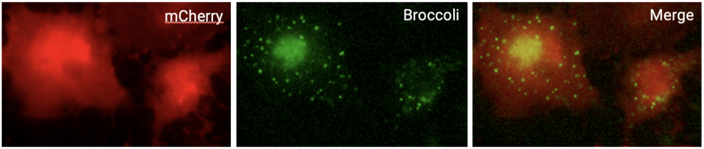
Figure 2. For single-molecule imaging, COS7 cells were transfected with a construct expressing mCherry-24xBroccoli™ transcripts and imaged in the presence 10 µM BI. Images of the same cells were acquired using TRITC and GFP filter sets to capture mCherry fluorescence (red) and Broccoli™ (green) signals. Mobile fluorescent puncta were detected in both the nuclei and cytosol of mCherry-expressing cells.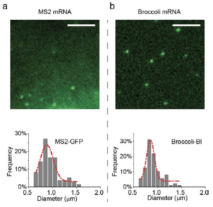
Figure 3. Live fluorescent mRNA puncta were imaged using COS7 cells expressing either A. the MS2-GFP system and B. the Broccoli™-BI system. The puncta size of Broccoil-tagged mRNA is similar to the puncta size of MS-tagged mRNA. Further, the puncta intensities of mRNA labeled from both systems exhibit a single Guassian distribution, consistent with the majority of these puncta reflecting single mRNA transcripts. The scale bars, are both 5 µm.Please contact us at for specific academic pricing.
More Details
-
- Properties
- Applications
- Reference
-
Overview
- Home
- Products
- Life Science
- Analytical Chromatography
- Assay Kits
- Biochemical Reagents
- Antibodies
- Antigens
- Enzymes
- Chemicals
- Proteins
- Liposomes
- Cell Culture
- Cell Analysis
- Cloning and Expression
- Nucleic Acid Synthesis
- Nucleic Acid Extraction
- Nucleic Acid Electrophoresis
- PCR
- Protein Gel Electrophoresis
- Protein Biochemistry
- Nanoparticles
- Glycan Analysis
- Immunohistochemistry (IHC)
- Microbiology
- Biospecimens
- Transfection Reagents
- Ion Exchange Resins
- Venoms
- Lab Solutions
- Lab Equipment
- Biosensors
- Flow Imaging Microscopes
- Fluorescence Quality Management Solutions
- Electrophoresis Systems
- Hematology Analyzers
- Isothermal Amplification Instruments
- Liquid Handling Equipment
- Luminometers
- Microfluidic Flow Control Systems
- Next-generation Sequencing Systems
- Nucleic Acid Purification Instruments
- Raman Spectroscopy Systems
- Sample Tracking Systems
- Spectrometer Accessories
- Syringe Pumps
- Thawing and Warming Systems
- Tissue Dissociation Systems
- Water Purification Systems
- Western Blotting Equipment
- General Laboratory Equipment
- Clinical Chemistry Analyzers
- Amerigo Scientific Instrument
- Automated Cell Selection Systems
- Automated Nucleic Acid Extraction Instruments
- Balances, Scales and Weighing
- Biolayer Interferometry Instruments
- Bioprocess Filtration Equipment
- Bioprocess Systems and Accessories
- Bioreactors and Fermenters
- Cappers and Decappers
- Cell Thawing Instruments
- Chromatography
- Data Loggers
- Droplet Generators
- Dry Baths
- Flow Cytometers
- Histology Laboratory Equipment
- Lab Ovens and Furnaces
- Lab Refrigeration Equipment
- Lab Shakers and Mixers
- Laboratory Centrifuges
- Laboratory Evaporators
- Laboratory Hoods
- Laboratory Incubators
- Laboratory Microscopes
- Laboratory Mills
- Laboratory Pumps
- Liquid Handling
- Live Cell Imaging Systems
- Microbiology Equipment
- Microscopy Flow Cells
- Mutation Breeding Instruments
- Oligonucleotide Synthesis Devices
- Particle Counters
- Plasma Surface Treatment Equipment
- Presses and Pellet Dies
- Surface Plasmon Resonance Instruments
- Ultrasonic Cleaners
- UV Disinfection Systems
- VOC Analysis Equipment
- VPHP Sterilization Systems
- Water Baths
- Lab Equipment
- Diagnostics & Clinical
- Applied Science
- Life Science
- Services
- About Us
- Contact Us


 .
.


