
-
-
Overview
-
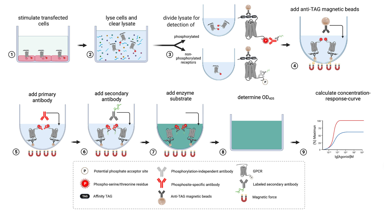
The pT354-D1 Phosphorylation Assay Kit allows determination of D1 Phosphorylation in 96-well plates without the need for Western blot analysis.
Please contact us at for specific academic pricing.
Background
Analysis of agonist-driven GPCR phosphorylation provides insights into the receptor activation state and ligand pharmacology. We offers Phosphorylation Assay kits based on the first-in-class immunoassay for the quantitative assessment of GPCR phosphorylation. The assay can be performed entirely in multiwell cell culture plates, thus eliminating the need for Western blot analysis. The assay involves immunoprecipitation of affinity-tagged receptors using magnetic beads followed by detection using Premium Phosphosite-Specific Antibodies as phospho-biosensors. The Receptor Phosphorylation Assay allows quantitative determination of GPCR phosphorylation particular when total receptor is determined in parallel using Non-Phospho Antibodies, which are included in each kit. The Receptor Phosphorylation Assay can be performed using transiently or stably transfected cells. This assay protocol has been optimized for transiently transfected HEK293 cells in 96-well format using plasmids expressing 3xHA-tagged GPCR constructs. It can be adapted to other affinity tags such as FLAG, Myc, His or GFP. The assay can be performed manually or fully automated in medium- to high-throughput mode. The Phosphorylation Assay Kit provides a fast, robust and reliable solution, fulfilling the requirements for extensive application in academic and pharmaceutical research.
More Details
-
- Properties
- Applications
-
Overview
- Home
- Products
- Life Science
- Analytical Chromatography
- Assay Kits
- Biochemical Reagents
- Antibodies
- Antigens
- Enzymes
- Chemicals
- Proteins
- Liposomes
- Cell Culture
- Cell Analysis
- Cloning and Expression
- Nucleic Acid Synthesis
- Nucleic Acid Extraction
- Nucleic Acid Electrophoresis
- PCR
- Protein Gel Electrophoresis
- Protein Biochemistry
- Nanoparticles
- Glycan Analysis
- Immunohistochemistry (IHC)
- Microbiology
- Biospecimens
- Transfection Reagents
- Ion Exchange Resins
- Venoms
- Lab Solutions
- Lab Equipment
- Biosensors
- Flow Imaging Microscopes
- Fluorescence Quality Management Solutions
- Electrophoresis Systems
- Hematology Analyzers
- Isothermal Amplification Instruments
- Liquid Handling Equipment
- Luminometers
- Microfluidic Flow Control Systems
- Next-generation Sequencing Systems
- Nucleic Acid Purification Instruments
- Raman Spectroscopy Systems
- Sample Tracking Systems
- Spectrometer Accessories
- Syringe Pumps
- Thawing and Warming Systems
- Tissue Dissociation Systems
- Water Purification Systems
- Western Blotting Equipment
- General Laboratory Equipment
- Clinical Chemistry Analyzers
- Amerigo Scientific Instrument
- Automated Cell Selection Systems
- Automated Nucleic Acid Extraction Instruments
- Balances, Scales and Weighing
- Biolayer Interferometry Instruments
- Bioprocess Filtration Equipment
- Bioprocess Systems and Accessories
- Bioreactors and Fermenters
- Cappers and Decappers
- Cell Thawing Instruments
- Chromatography
- Data Loggers
- Droplet Generators
- Dry Baths
- Flow Cytometers
- Histology Laboratory Equipment
- Lab Ovens and Furnaces
- Lab Refrigeration Equipment
- Lab Shakers and Mixers
- Laboratory Centrifuges
- Laboratory Evaporators
- Laboratory Hoods
- Laboratory Incubators
- Laboratory Microscopes
- Laboratory Mills
- Laboratory Pumps
- Liquid Handling
- Live Cell Imaging Systems
- Microbiology Equipment
- Microscopy Flow Cells
- Mutation Breeding Instruments
- Oligonucleotide Synthesis Devices
- Particle Counters
- Plasma Surface Treatment Equipment
- Presses and Pellet Dies
- Surface Plasmon Resonance Instruments
- Ultrasonic Cleaners
- UV Disinfection Systems
- VOC Analysis Equipment
- VPHP Sterilization Systems
- Water Baths
- Lab Equipment
- Diagnostics & Clinical
- Applied Science
- Life Science
- Services
- About Us
- Contact Us


 .
.


